
Related Images


Download:
| Tiny | 170x128 | View | Download |
| Small | 341x256 | View | Download |
| Medium | 682x512 | View | Download |
| Large | 1365x1024 | View | Download |
| Original png | 1440x1080 | View | Download |
| Original as jpg | 1440x1080 | View | Download |
This image was acquired from
wikimedia. It was marked as Public Domain or CC0 and is free to use. To verify, go to the source and check the information there.
Looking for more info about this image?
Try a Google Reverse Image Search for it.
Try a Google Reverse Image Search for it.
Keywords from Image Description:
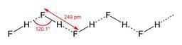
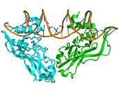
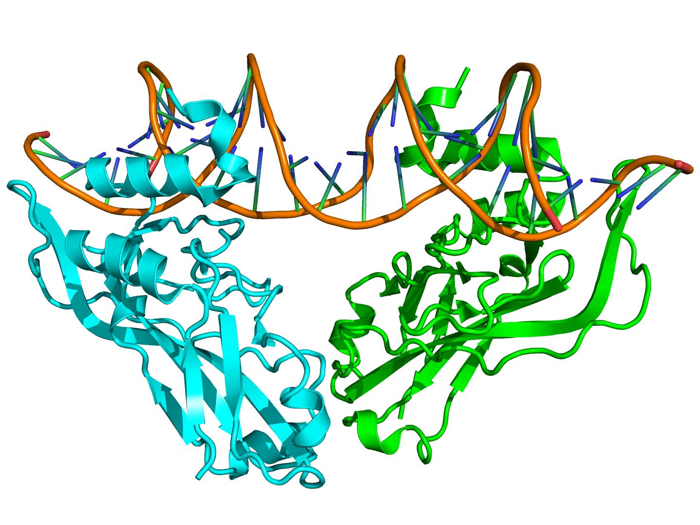
HF. Crystallographic structure of the TBX protein dimer cyan and green complexed with DNA brown based on the hf coordinates selfmade using PyMol Boghog DNAbinding proteins