
Related Images


Download:
| Tiny | 203x128 | View | Download |
| Small | 407x256 | View | Download |
| Medium | 814x512 | View | Download |
| Large | 1628x1024 | View | Download |
| Original png | 2391x1503 | View | Download |
| Original as jpg | 2391x1503 | View | Download |
This image was acquired from
wikimedia. It was marked as Public Domain or CC0 and is free to use. To verify, go to the source and check the information there.
Looking for more info about this image?
Try a Google Reverse Image Search for it.
Try a Google Reverse Image Search for it.
Keywords from Image Description:
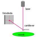
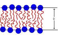
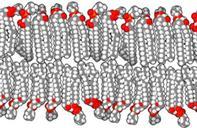
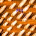
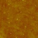
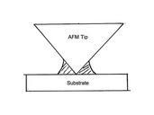
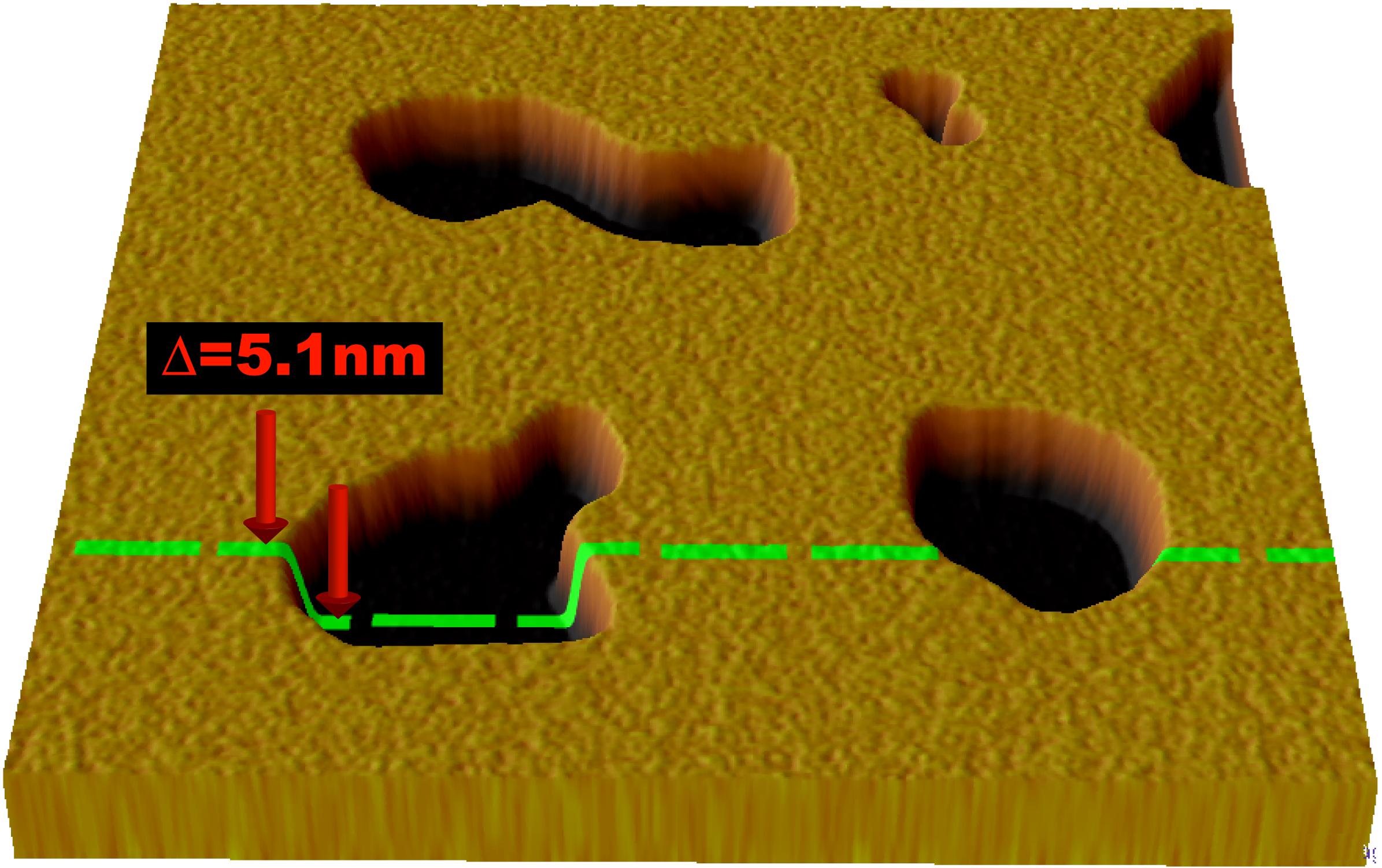
Bilayer AFM schematic. en Illustration of typical result from an AFM scan of supported lipid bilayer The bilayer shown here has several defects which appear as pits By measuring the distance from the top of the bilayer to the substrate it is possible to determine the thickness of the bilayer own MDougM Ultrastructure diagrams Cell