Related Images

Download:
| Tiny | 174x128 | View | Download |
| Small | 349x256 | View | Download |
| Medium | 698x512 | View | Download |
| Large | 1397x1024 | View | Download |
| Original png | 1700x1246 | View | Download |
| Original as jpg | 1700x1246 | View | Download |
This image was acquired from
wikimedia. It was marked as Public Domain or CC0 and is free to use. To verify, go to the source and check the information there.
Looking for more info about this image?
Try a Google Reverse Image Search for it.
Try a Google Reverse Image Search for it.
Keywords from Image Description:
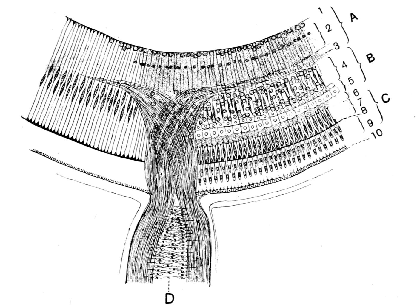
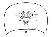
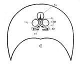
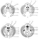
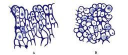
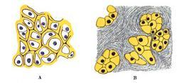
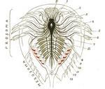
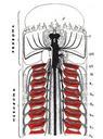
Origin of Vertebrates Fig . Fig Retina and Optic Nerve of Petromyzon After Mller and Langerhans On the left side the Mllerian fibres and pigmentepithelium are represented alone The retina is divided into an epithelial part the layer of visual rodcells and neurodermal or cerebral part which is formed of the ganglion of the optic