Related Images


Download:
| Tiny | 128x128 | View | Download |
| Small | 256x256 | View | Download |
| Medium | 512x512 | View | Download |
| Original png | 1000x1000 | View | Download |
| Original as jpg | 1000x1000 | View | Download |
This image was acquired from
wikimedia. It was marked as Public Domain or CC0 and is free to use. To verify, go to the source and check the information there.
Looking for more info about this image?
Try a Google Reverse Image Search for it.
Try a Google Reverse Image Search for it.
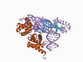
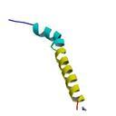
Keywords from Image Description:
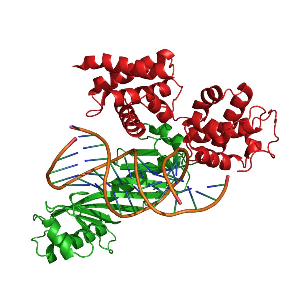
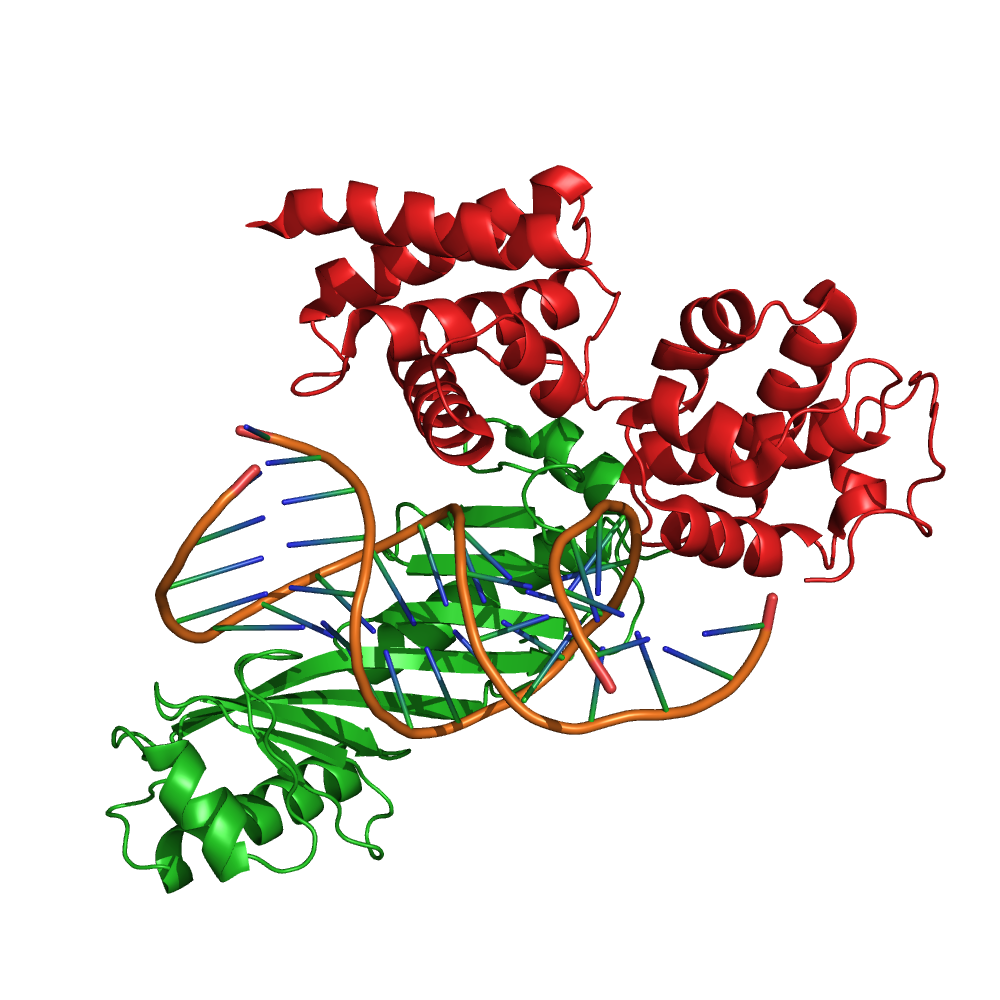
PDB rendering of TFIIB core based on CB. Uploaded with en wp UW marker Structure of the core domain of TFIIB red in complex with adenoviral major late promoter and TBP green rcsb org do structureId CB Tsai Sigler rcsb org do generalinformation html Tsai Sigler