
Related Images


Download:
| Tiny | 170x128 | View | Download |
| Small | 341x256 | View | Download |
| Medium | 682x512 | View | Download |
| Original | 1280x960 | View | Download |
This image was acquired from
wikimedia. It was marked as Public Domain or CC0 and is free to use. To verify, go to the source and check the information there.
Looking for more info about this image?
Try a Google Reverse Image Search for it.
Try a Google Reverse Image Search for it.
Keywords from Image Description:
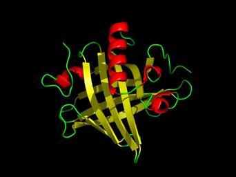
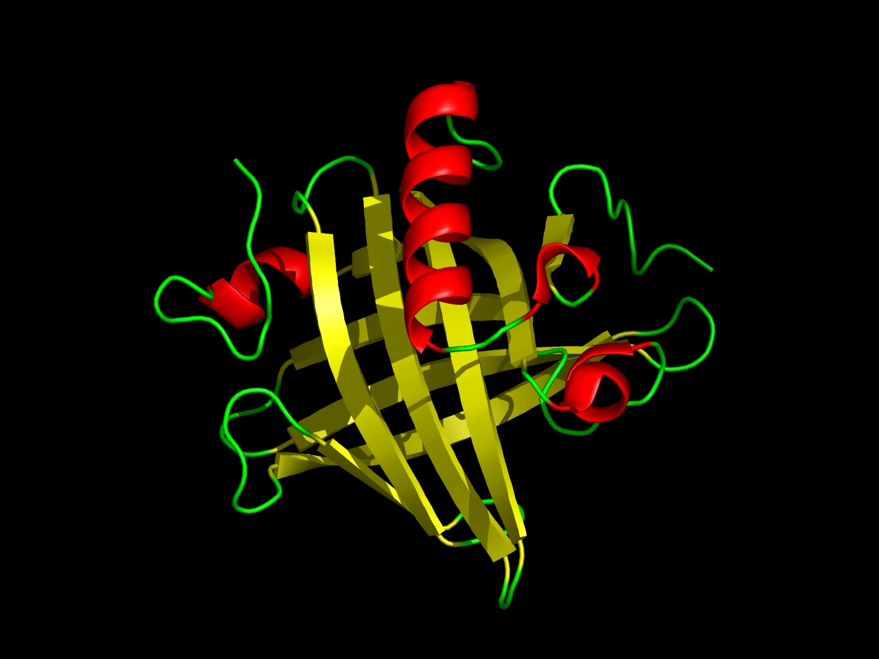
Mup PDB i. en February Tertiary structure of mouse major urinary protein Resolved from rcsb org do structureId PDB The protein has eight beta sheets yellow arranged in beta barrel open at one end with alpha helices red at both the amino and carboxyl termini created this work entirely by myself using dkfz de php AISMIG Rockpocket